The Operation; Part 1
- Transcript
<v Man 1>8 Alive. Pacific Mountain Network Special. Major funding for The Operation has been provided by a grant from the International Heart Foundation with additional funding provided by Bard cardiopulmonary division of C.R. Bard Incorporated, Bio-Medicus Incorporated, and Shiley Incoporated Irvine, California. [music plays] <v Dr. Edward B. Diethrich>Good evening. Over the past 10 years, we have used television extensively to educate nurses, technicians and physicians on the latest techniques of open heart surgery. There is a ?inaudible? policy of service includes a commitment to public education as well. Most noteworthy has been our use of the media to advance the concept of early recognition and prevention of heart disease. Indeed, is our belief that the appropriate corrective and preventive measures are taken. The type of open heart procedure you're about to see can often be prevented. Since the schedulin' of this live heart operation, numerous people have asked, why would we participate in this public information program? In fact, there's been some skepticism and even criticism by both lay and professional colleagues. Let me see if I can clarify that question before we go in the operating room. First, it is my feeling that people are less likely to fear what they know and understand. It is the unknown that scares us. We will show you tonight a coronary artery bypass procedure on a 62 year old gentleman who has suffered a small heart attack but suffers more now from chest pain due to insufficient blood supply to the heart muscle. That educational experience should relieve anxiety about the most common heart operation we do in the United States today. In fact, it may even sensitize you to the point that you will do something about preventing your own heart attack in the future. Second, there is a misconception that heart surgery cures heart disease. The operation is only one component of the treatment program for the patient. We want to explain risk factors of coronary ?artery? disease, prevention techniques, and the cardiac conditioning that closes the loop following the coronary bypass operation. The goal of heart care utilizing cardiac surgery is prolongation of a good quality of life. Finally, I want you to see the procedure because each of you has a heart within your body. It is an amazing organ, beautiful in its function, its character, and its capability. You deserve to see the beating heart at least once in your lifetime. And why live? Because life is alive and that is the way you should experience it. Please join me now with our surgical team for the operation.
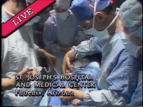
<v Rick Dolly>I'm Rick Dolly, KAET's science editor and your host for The Operation. With me in our Arizona State University studio is Dr. Sam Kinard, a cardiologist and director of the cardiac catheterization lab at the St. Joseph's Hospital and Medical Center in Phenix, Arizona. The patient is Mr. Bernard Schuler. We shall return to the specifics of his case after Dr. Edward B. Diethrich, medical director of the Arizona Heart Institute, introduces the surgical team at the St. Joseph's Hospital and Medical Center Operating Theater.
<v Speaker>Dr. Diethrich, good evening in the studio. Can you hear us all right. Loud and clear. Good. We'd like to take you on a quick tour of the operating room before we start the actual operation. <v Speaker>I want to introduce first Dr Nelson Dias, who's at the head of the table. Dr. Dias will be giving anesthetic to saving at your dad's. Would you get an idea about your function this evening or this procedure? <v Speaker>Heart surgery is very much like out of surgery, except that during the actual work on the delicate work on the heart arteries, the heart and the lungs are stopped there. The lungs are not ventilated. And the and the heart is not beating. <v Speaker>This is possible only because of the heart lung machine that is across the room over here. This is this diverse, the blood from the venous side, from the tissues to intercepted, taken and oxygenated, returned to the patients with oxygen.
<v Speaker>Good. Let's take a look at that. If we can swing the camera out. We'll show the heart lung machine. And Gary Mandele, who is our candidate, former perfusionist Gary. Can you just give an overview of the heart lung machine and just show what it's going to do to see what it's going to be doing this evening as the blood's going to come back to me? <v Speaker>It will be oxygenated and then I'll send it back to the patient. The blood that we send back to the patient will be fresh with oxygen and you will see the difference later in the procedure. <v Speaker>The final area I'd like to show you is the back table where we have the instruments assembled. We can show you a variety of instruments. And later on, I'll ask one of our scrub nurses to describe some of these instruments and how we use them. Got to kind of the first part of any operation is the incision. And we're in a position now where I'd like to show you the incision. This is the only part of the operating procedure which may make you a little bit queasy. And I would say to the viewing audience, if you feel that you're going to be bothered by the incision and a small amount of blood at this time, probably you should turn your head on the screen. Once we've entered the chest, the remainder of the procedure, I think we'll find to be beautiful and very instructive. <v Speaker>The incision in an open heart procedure is begun at the upper end of the sternum and continues down to a level which is just below the breastbone. If each of the viewing audience is actually feel their chest with their hand, they can feel this best ball. We'll use this electrode cordery now to come through the soft tissue and go down to the rest below, which we call the sternum. Now, for a few minutes, Doctor kind of will be coagulating the blood vessel. This might be an ideal time for you. Describe the patient that we operating on this evening. Thank you.
<v Speaker>To the patient we have today is a 62 year old man who had, as Dr. Dietrich said, a mild heart attack in 1977. He was well until December when he developed angina pectoris or chest pain related to exertion. He was treated medically, but his pain did not improve. Therefore, he sought further advice. He has risk factors of having smoked. His father died of hardening of the arteries. His cholesterol is at the higher limits of normal. His electrocardiogram shows changes related to a small scar. And on his treadmill, he had changes that we associate with lack of blood supply to his heart. <v Speaker>The artery Graham demonstrates occlusions. Neuro occlusion of two vessels. The circumflex Karner artery, which supplies about 50 percent of his heart, is about 90 percent occluded or stopped up with the plaque. The anterior descending is about 75 percent stopped up with this plaque. So this is the setting of our patients this evening.
<v Speaker>The surgical procedure you will be viewing is one of many alternative strategies for improving the odds against a heart attack and for relieving the painful symptoms associated with heart disease. Mr. Sures case, as in all patients who have had a heart attack, the problem is with the coronary arteries. Doctor, kind of. I'd like you to explain exactly what we're looking at. <v Speaker>This is the external anatomy of the heart, and you'll be able to see this later in the operation, but not quite as well, probably. You see the signs to identify the coronary arteries which arise from the aorta. The left karner artery here divides into the circumflex, which goes around and between the left atrium or the left upper chamber and the left ventricle and the anterior descending courses along the front part of the heart. The right goes around the opposite side of the heart from the circumflex. The pulmonary artery, which is the artery to the lungs, covers up the base of the left main coronary artery so that you will not be able to see it too well. <v Speaker>And if we could interrupt you for a moment now we're progressing along with the operation. I would like to have you see the opening of the sternum.
<v Speaker>This is a Sternhell sore. It's a special instrument we use to open the breast ball. You can see it is an actuating shot. You can put your finger right on the. <v Speaker>I won't even cut it. But once it presses up against the sternum or the breastbone, it will crack the sternum very easily. <v Speaker>Just placed the sternum sore at the upper end. <v Speaker>You can see how rapidly we can split that sternum. Now we use a little bone wax on the edge of the sternum here because there's often some tiny little vessels on the edge of the bone. Whenever we cut the bone, I think you can see that pretty clearly. I'm sorry to interrupt you. You may go back to your assistant. I didn't want you to miss the opening of a stern. <v Speaker>What we were just seeing and what you were just discussing, Doctor, kind of was, of course, the anatomy of a normal heart. Now, if one or more of those coronary arteries that we were looking at in the diagram should fail, then the heart muscle will starve and eventually die, a condition called a myocardial infarction or heart attack. Doctor, I'd like you to explain just how those coronary arteries, in fact, do fail to perform their function. <v Speaker>Well, the blood, of course, passes from the left ventricle out into the aorta and down into the small karner arteries, which supply the heart with blood. If a plaque forms within these vessels, then this progressively increases over a period of life of a lifetime, probably. And if it reaches the point that there's not enough flow through the vessel, then we have what we call a schema or lack of blood supply to the heart. If this progresses, so that is so severe that the muscle does not get enough blood to maintain life, then we have death of heart muscle or a heart attack or a myocardial infarction. If something is done prior to myocardial infarction, then the muscle can be saved. If, on the other hand, myocardial infarction ensues, then this is a an event which cannot be replaced because all muscle die. Once the muscle dies, it cannot be brought back. If one places a bypass or a vessel or conduit which goes around the plaque into the artery, then one can bring more blood supply to this artery and improve the circulation and relieve the situation of what we call a schema. And that's what we're talking about tonight with this patient.
<v Speaker>Well, now, in order to prove that those obstructions are there, we do a test called an arteriogram or angiogram. In that case, a thin, flexible tube called a catheter is pushed up a major artery from the leg or the arm to the level of the heart.
<v Speaker>Can I interrupt you one more second? Surely this is an important point of the operation now. I don't want to miss this. This is the pericardial sac, the sac, which surrounds the heart. We're going to open this sac and that will be the first gear we have of the heart, which is right below the sac, encompassed and surrounded in its entirety. Most people believe that the heart lies fairly far on the left side of the chest. <v Speaker>But as you can see here, as we enter the heart itself is beginning sort of at the center of the chest, and that extends into the left side to extend our incision now up over the order. <v Speaker>And the moment we'll show that anatomy, as you just heard in your diagram, you can then extend the incision down toward the foot. <v Speaker>Now for your orientation, a patient first is going to my right side. <v Speaker>Down this way, the patient's head is to my left side up here. And we'll just point that out several times as we go, because it's a little confusing in terms of the orientation. Now, this is the first year you have of the heart. We'll open our external spreader a little more. We can place a silk suture along the pericardial sac and it will give us a good view here.
<v Speaker>The arson in the order here is the acid in your. Now, this is a large vessel that comes from the heart. <v Speaker>The left side, the heart and supplies the blood through the entire body. First of all, going after the brains, then to the upper extremities. And then finally down to the lower extremity. <v Speaker>Ascending aorta, as we discussed, is the big vessel, which from which will rise also the coroner arteries, which, of course, pass along the surface of the heart, which we will see in a minute. <v Speaker>Now, what we're doing at this particular point that, Kyra, is cutting the connective tissue, which overlies the order we need to have a enough area of the order here so that we can do what we call the cannulation.
<v Speaker>There are occasionally some small little blood vessels up here, and you can see how we use electric Connery to cauterize those blood vessels. <v Speaker>We can also get a little more freedom of the order by cutting this connective tissue. This is loose, fat, connective tissue about the normal amount that we see on the overlying the order. And as we do this, you get a very good view then of what the action in the order actually looks like. It's. Now, in order to connect the patient to the heart lung machine and what we have to do is place a cannula or what we call a two really here in the S in the order, in order to do that, we will place a small first string suture. You see, this is a monofilament suture. It's quite fine in its nature. And it slides through the artery very smoothly. And it's through those purse strings, searcher, that we'll place the formula are the two which will bring the blood from the pump action back to the patient. <v Speaker>Now, as soon as I push those purse strings and I think it would be a good idea for us to have Gary Mandele back at the heart lung machine show what the circuit is going to be as the blood comes from the patient. So as we finish putting this suture and if we can switch the camera back, I'd like to have that kind of maybe on the screen you can explain the circuit.
<v Speaker>And then also very can show them lie here in the operating room what the heart lung machine looks like. <v Speaker>Yes. This is a demonstration to pump to oxygen later at at the was at the top of the film. And the blood passes, of course, around from the heart into the heart lung machine where it's oxygenated, brought back into the aorta after his oxygenated in the heart lung machine. Gary, you might want to point out the tubes that do this. <v Speaker>The larger one right here is the one coming from the patient. That's what we call our venous line from there. It'll drain back into the oxygen later. It'll be oxygenated in this column. It'll also be cooled in this column. The oxygen will push the up the blood over the top and it'll drain into what we call our arterial reservoir. <v Speaker>And all that is, is a storing chamber for all the fresh, oxygenated blood from here. It'll be pulled over to a biomedical arts center, difficult vortex pump, and it'll be pushed out through an arterial filter to the patient.
<v Speaker>We're ready now to give a heparin. Dr. Dyas has heparin at the head of the table, and he's going to in a moment. Should that happen and doctor. Yes. Go ahead and put the. Happened in and then we will. <v Speaker>I think we can show that on the camera going in. We should mention that the purpose of heparin is to prevent the blood from clotting within the heart lung machine. <v Speaker>Once a heparin is circulated around the entire cardiovascular system, we'll go ahead and put this cannula into the acid in your order. And this might be a good time. Also, Dr. Dayas forced to show the pressure on the screen and explain the electrocardiogram and pressure as soon as you put this can Tanyalee. And I think we can go ahead and do that now. Is there enough time for the heparin to circulate, replace the cannulation? <v Speaker>And then as we're securing this, we'll go ahead and show the screen up there. Dr. Dias, would you please explain what those lines are and how you're going to watch them and what they mean during the procedure?
<v Speaker>This is the blood pressure tracing right here. The haziq wave we read would read this blood pressure approximately one hundred and tan over 60, which is very normal. This is a central venous pressure line. It gives us an idea of the blood volume balance in the patient. These two leaders up here are electric party graphic leads that tell us something about the electrical activity of the heart. <v Speaker>We're ready to connect this tubing up to the line, which Feary has passed up to us. Now, it's very important at this point that there be no air in this particular line. So we'll let a little this blood come out the tap to be sure there's no air and then connect it directly to the perfusion line, which will be coming from the. <v Speaker>We should point out that this is the first connection made to the pump and will be the return flow of oxygen, oxygenated blood. It is the line that is substituting for the main pump up the heart. That is correct.
<v Speaker>It will connect. Now, the area on the heart. This is the right record appendage. I think you can see that fairly clearly. We'll place a partial occluding clamp on that. And then we will take a soak suture and go around that. A record appendage. It's through the Sorich Appendage that we will place the cannula or the tube, which will then drain the blood from the body back to the heart lung machine. As you saw on the diagram. <v Speaker>And Gary explained, the right irregular appendage is essentially an extension, is it of the right oracle or right atrium? <v Speaker>Yes. Right. Upper Chamber of the heart, which receives all the blood from the body. <v Speaker>Into the right. Our oracle drains the superior vena cava and the inferior vena cava.
<v Speaker>And of course, he must get all the blood from all of the body so he has a cannula which goes into the inferior vena cava and gets the blood from there. And then it has holes proximal to this which suck out the blood that comes in there. <v Speaker>Dr. Carter, right inside the the irregular appendage, you can see that if we take the clamp off it, we're looking right inside the heart. And we have this special kanya, these cannulas are made by the Abad Corporation. And this particular one, as you can see, has holes at the tip that holds the middle portion. So we can drain the inferior vena cava and also the superior through the right atrium. With this one cannula already guided in, I'll put my hand bottom behind and that will slip then right into the in fear being the case. <v Speaker>And again, what we're doing here is we're making the second connection to the pump. This is the flow that comes from the body that needs to be oxygenated and goes into the pump for the oxygenation.
<v Speaker>Correct. This would normally go through the lungs, but the lungs are to be separated from the heart and the heart lung machine is the lungs. <v Speaker>And once this connection is made, the circuit essentially is complete. And the heart is bypassed. Correct. <v Speaker>Once again, we'll fill this tubing. You see that blood? Now you notice the difference in color. I think the camera can pick that up. This this is rather dark blood because it's coming from the right side of the heart, is desaturated and does not have a great deal of action in it. <v Speaker>Now, if we can turn the cameras, I'd like to have Gary show the flow of blood from the body. We keep the clamps off and put the patient on a heart lung machine. We'll remove these clamps now and you'll see the flow of blood coming from the body. And Gary, why did you pick it up and describe the circuit going through? <v Speaker>Blood is now oxygenated at this point. It's drained into the arterial reservoir.
<v Speaker>And now it's being returned to the patient. It's going through the bio medicus. Through a filter. And then on up to the patient, I noticed the difference in the two colors of blood. The desaturated blood on your left and your arterial blood. Full of oxygen on the right hand side. <v Speaker>Again, desaturated, meaning depleted in oxygen, that blood, which is being returned to the heart from the rest of the body. <v Speaker>More on the heart lung machine now. Now, that's kind of what we'll do is place this clamp across the pass in an aorta. This trocar will go into the assignee order, not through this trocar. They will place a very cold solution. It's five degrees centigrade and that solution also contains a large amount of potassium. You can see that. Now, let me lift the heart up. You'll see the heart is beating so very good through the heart. But notice how slow it will begin to beat. Now, in a minute, it's going to be completely stopped. That's because we're now cooling with the cardioplegia solution. Adak, Dias forwork doing this. You might like to describe what is in there. How do you plead the solution? <v Speaker>This is the cardioplegia solution, and this bag, like Dr. Dietrich says, it has potassium plasm eight.
<v Speaker>It's very cold. The cold is to cool the heart so that it requires very little oxygen. While it's not beating. There's a Tascam stops the heart from beating and also for terrorism. <v Speaker>Let's get to it. Let's go to. <v Speaker>I should mention that the heart was one lapse because all of the blood now is going through the heart lung machine. So they look different than it does when we first got here. <v Speaker>And if we were now to look at the blood pressure on that monitor that we saw originally in The Shot, we would not be seeing the typical blood pressure that isolates from a high to a low to a high, a low risk, as you saw on the heart lung machine there as a roller pump which circulates the blood. <v Speaker>Now, rather than the contraction and relaxation of the heart so that instead of a sudden rise and drop in pressure, there is now just an oscillating wave of the blood pressure on the machine on the oscilloscope. And we'll see that later. <v Speaker>So essentially, the heart lung machine is massaging the blood through those plastic tubes. That's correct. We might point out for the viewing audience that most of the temperatures you'll hear tonight are in degrees centigrade or Celsius, not in Fahrenheit. That five degrees Celsius is a lot colder. It's not quite as cold as it sounds. I think that calculates in Fahrenheit to about 41 degrees. Still quite cold, but enough to stop the heart from being that plus potassium. But the potassium salt also does the same.
<v Speaker>The cold also protects the heart against a schema or lack of flow during the period that the operation is being done. <v Speaker>I might mention that this patient has a very large circumflex coronary artery, which is the artery which we saw. That is in the groove between the left upper chamber and the left lower chamber. And this is the one of the two arteries that need to be bypassed. <v Speaker>There was some discussion prior as to whether one would be able to do the connection directly to the circumflex, or one might have to go to some of the branches of the circumflex. And of course, he is Dr. Dietrich is looking around now and making this decision. I assume, as he is looking far back at the back part of the heart in the avy groove or the grube between the left upper and left lower chamber.
<v Speaker>This is a difficult area to get to because it is so high at the base of the heart and the base of the heart is connected there as opposed to the apex of the heart, which you could see he could raise out of the pericardial sac or the sac within which the heart risks peritta. <v Speaker>Kind of what we're doing now. We have found the main artery that was blocked coming off. We've also found a poster descending, coming off of the circumflex and we'll bypass. I think those two arteries will probably do a jump graft from one to the other. The one that's higher up is here, a second branch here coming off the to be sunny. So we'll probably jump both of those arteries. We're also looking around, though, to see if there are any other areas where we might want to bypass and why we're doing this. <v Speaker>It might be a good time for us to demonstrate some of the instruments that we use because we're going to be using several of these now. Now, I asked Patti Thompson if she'll show the back table and show us some of these.
<v Speaker>We have over 400 instruments on the back table, over half of them. Our instruments used in all types of surgery. <v Speaker>We have retractors for exposure. Needle holders for suturing, forceps for grafting, feathers for bisecting and hemostat for clamping. Here are two types of hemostat. Its function is the same, although they look very different. This is for very tiny vessel and shallow tissue, sensitive to occlude larger vessels and deeper tissue. The rest of the instruments are specialized for vascular and cardiac surgery. Most of the vascular clamps are specifically designed to totally occlude or partially include veins and arteries throughout the body. Imagine that this is a damaged artery. You can see that by totally including it on both sides. Two, seven years. And that's the blood flow and blood loss is eliminated. And the surgeon can then repair the artery in a bloodless field at other times to the patients and manage to partially occlude the vessel and repair it without interference, interfering with blood flow. The other specialized instruments we have for bypass surgery consist of these very delicate scissors. Dr. Dietrich designed Maeder. <v Speaker>I'm just one second. You could back any moment, but I would like to show the vein that's been removed by Dr. Bower's, you and Dr. Selke on the back and you demonstrate that vein because we need to pass that up now. We're going to begin the suturing if we can show the vein on the back table. Can you see the vein that. Yes, we see it. Well, good. I will have that vein passed up to us now, because that's an extra vein that's been taken from the leg. We call a superficial vein and we'll pass that up here into the operating side so that we can begin the soul of the vein onto the coronary artery.
<v Speaker>When he says extra, he means that there are other veins which do the same job as this vein. So it can be removed without problems. <v Speaker>And in fact, in earlier discussions that we have had before the show, you pointed out that uh common procedure called vein stripping routinely makes use of the removal of the south and ?inaudible?. <v Speaker>That's right. And at times that is a problem in a patient who has had veins dripping in the past and needs a coronary bypass when he gets older, he or she. <v Speaker>The veins have valves in them which prevent the flow of blood back down the leg and one is standing up. And therefore, these are one way valves. And so the vein must be turned around so that the blood flows uh in the same way that it would in the leg. But uh in a proper way, so that the valve does not obstruct flow.
<v Speaker>The small branches of the vein, of course, have to be tied off so that there's no bleeding from the vein. And you'll probably be able to see some black marks along the vein where they have put silk suture uh on the branches of the vein to th- to prevent bleeding. <v Speaker>Now, in this exceedingly tight shot where the end of the graft vein is being sutured directly to the coronary artery, they're using a fine, I assume, monofilament synthetic. <v Speaker>That is correct. <v Speaker>Uh material.
<v Speaker>The uh- <v Speaker>?inaudible? <v Speaker>As I said before, this is a, as you can see, a a difficult position to uh get to because it's on the back part of the heart and at the base of the heart. Uh usually the anastamosis are connection of the vein to the coronary artery is made first and then the other connection of the vein to the aorta is made second and one can check after one makes the connection to the coroner artery with a fluid to see if there's leaking uh around the uh connection where the sutures are placed and then put more sutures if it's necessary. After the patient comes off the heart lung machine and the clotting is changed is the heparin is reversed. Then one looks again to make sure there's not bleeding because then clots form in the needle hold. And this seals off the connection of the vein to the coroner artery. And the connection of the vein to the aorta. <v Speaker>We're proceeding very nicely with his side. As you said, we call this in anastamosis. We have selected one of these branches coming off the circumflex and we have got about three quarters of the way around the artery taking the vein down to the artery so that the vein after we've completed this will actually act as a detering artery or new artery for the patient.
<v Speaker>It's a little hard for you to see, perhaps because you are looking sort of over the heart. We'll have a better view of the coronary artery in a few moments when we do one or the other. And that's the most used to the answer descending, can't we? We're doing this. There's a lot, of course, going on in the operating room with the rest of the team. <v Speaker>Dr. Dias is monitoring the blood gases, the salts and the hemoglobin and Medicaid and a lot of other things that I'd like to have him demonstrate for you as we're doing this suturing technique. I did ask for your show, the audience, what you're doing there. <v Speaker>You look back at the screen, you can see it's much different from what it was before we went on the bypass. This is the arterial pressure line now. It's I mean, blood pressure. It's about seventy five, which is just where we want it to be. The reason it's flat is because the roller head's, instead of having a parts of town flow, has a continuous flow. This, again, is a central Munish pressure line. The EKG leads are both straight lines because the heart is stopped and not meeting. We can go to the blood gas monitor. These are blood gases that were drawn before the patient went into the room. There's several different tests on each one of these. You can see here this is the time at five, 30 p.m. this afternoon, six, 14, six thirty two. And then there's various things here. The P.H., which is one of our important test, indicates the patient's acid base balance. And here it's seven point four to seven point four four, seven point four five. All these values are within normal limits and we'll try to keep them there for the entire operation. This is the oxygen tension, the partial pressure of oxygen. You can see it was 76, a little bit on the low side. This was without oxygen in the holding area. Both of these 135, 140 forty or after we started the anesthesia. So he was being ventilated and, well, oxygenated. This is the back carbon.
<v Speaker>At the next level is the hemoglobin level. And you can see it. It is fifteen point six, fourteen point four normal or perhaps just a little above normal. This is a potassium ion and it's within the normal.
<v Speaker>Gary, you might tell us we didn't talk about cooling the patient. Can we go back and show on the heart lung machine how Gary's my attempt here? And Gary, would you give us an idea of what you planned to do in terms of monarch? Lower the pressure and the capture. <v Speaker>As we are on the heart lung machine, we're going to cool down to twenty five degrees Celsius, which in Fahrenheit is about 77 degrees, and we'll leave it at that temperature until all the distal grafts are attached to the heart. <v Speaker>And what is the temperature right now? Right now we are 25 or the 77 degrees Fahrenheit. <v Speaker>Now, not to kind of we completed one of these Anasta motions to the circumflex system. <v Speaker>We have another artery identified here. We are going to bypass. So we'll once again lower the vein into the pericardial sac and we will now. So this burn onto the artery just the same as we did the first one.
<v Speaker>I should mention, these are secondary branches of the circumflex, coronary artery circumflex being the large karner coronary artery going around in the avy groove or the group between the left upper left lower chambers. And these two secondary branches were the two identified at the time of the artier gram as the largest branches so that he was unable to get at the circumflex in the avy groove because it frequently is inaccessible there. And so it's easier to do this and ask the most as to these two branches than a single and asked the most is to try to do in a V groove. And these two branches together will allow enough blood to go back into the circumflex to furnish the entire distribution. The blood passes into these branches, both distally and proximally. In other words, it goes both ways so that it can go back into the circumflex as well as out into the distribution of a small branch. <v Speaker>We've heard the term pericardial sac used very often, I think you might describe for us what we're talking about, the pericardial sac is, as you saw when he opened first, you saw that there was sort of red looking tissue that had some blood on it.
<v Speaker>And then as he opened a sac, everything looks clean and without blood. This is a sac which holds the heart and allows it to move. And it has a little fluid within it, which sort of lubricates things as the heart moves around. Of course, any time one has any inflammation or, for instance, a heart attack which results in death of heart muscle, then one gets inflammation of the heart. And that results in inflammation in the pericardial sac. And so one may get actually connections between the pericardial sac and the heart muscle itself. Most of the tissues of the body which move are contained within a sac, for instance, the lungs are both contained within a sac as well. We're not at the present time with this incision. We have not opened the sacs that contain the lungs, but only the sac which contains the heart. <v Speaker>And so in brief, the pericardial sac has as its function o lubrication, if you will, or at least a sliding surface over which the heart can move in relation to the other organs around it. That's correct.
<v Speaker>We're finishing up now the anastamosis on the second, Harbury coming off the circumflex. We didn't show before. When we finish their anastamosis, there was a time Dr. Dyson explained what gases that we do, put some more cardioplegia solution directly through the vein graft itself that further calls the heart. We will demonstrate that in a moment here as we put the last ditch in. <v Speaker>Tradition asked Moses, who can raise the first being wrapped up, and you can see that, here's the second one. <v Speaker>And we'll put some solution on this doctor by. We'll shoot the cardioplegia solution in their. <v Speaker>And this way, we all show. So check to be sure there are no leaks. Now, the Anasta most, as you can see, that looks very good enough. She might point out that this post now will put several knots in this, because this monofilament searcher is a little on the slippery side. Want to be absolutely certain that it's. Right. And then we'll place a silver clip right where we do the Anasta Moses so that if ever in the future you need to examine the patient with another tangram when they're thirsty. You'll have the opportunity to know exactly where the UN estimates this is. I can see a little leaking right where the suture winning end. So we'll take one small suture here. Sometimes these small leaks don't show up until the heart starts to beat. And we have to put an extra stitch in at that time. Now, Paul Dursun is been holding the heart. She just let loose of that, you saw the heart go back into the pericardial sac to bring the vein graft around. And once again, we'll cut this vein to an appropriate length. The place, a hemostat, small mosquito, hemostat, and then. We'll tie this canula on to the vein so that we can periodically put more cardioplegia solution. Directly into the heart muscle. Now we've completed the two Anasta, Mosese or two Soren's through the circumflex system and we'll have a much better view of the heart here when we do the anterior descending. We'll place a lap behind the heart, will raise the heart right up. I think a very good view. Now, can you see the answer descending pretty well that we can see where it is. All right. I got to point that out for you. Here it comes, directly down the middle. Coming right down here, the middle. This is about the normal amount of fat that we would see in a heart on this side. We have the right ventricle on this side. We have a left ventricle. Now, in order to make the incision into the enter descending, we'll have to cut this fatty tissue, which is overline. And already, I think if the camera comes in very close, you'll have an idea of what the coronary artery looks like here.
<v Speaker>We'll just make a small incision with this number 11 blade.
<v Speaker>And then we have these special coronary scissors that Patty demonstrated a few moments ago. That's you will pass up this reverses. This is a unique kind of CERs or it goes the opposite direction, but it makes it very easy to make that cut into the coronary artery. Now, we'll place a retention suture on either side of the incision in the corner artery, and that will give you a very good view. Dick Williams is doing our overhead photography, and I think that he'll be able to. <v Speaker>Get a good shot of this and third, descending. We have an excellent shot. <v Speaker>Now, to give you some idea of what this size is. We'll pass a dilator. This dilator is a millimeter. I'd like to use about a millimeter and a half dilator holdfast, a millimeter and a half dilator into the corner territory. Now, if you watch, I can pass it up and I can feel at the top right there, you see a diagonal will go no further. That's because there's obstruction at that particular point. That's where the obstruction is in the corner. Now, the important thing is to be sure there's no friction down below. So we'll pass the dilator where we call distally or down. And you see there's no obstruction down here, passes very nicely. So we'll go ahead now. And so this final vein graft on to the anterior descending coronary artery will trim the vein to the appropriate length. We want to make a little larger incision in that just so it's exactly the length that we want. We'll suck the coronary artery because there's a little using a blood foam that nothing very much. And then we lower this vein graft right down on to the coronary artery. And this will begin then the Anasta most. This is a sewing between the vein graft in and through descending coronary artery. <v Speaker>We might point out at this time that in actual fact, there there were really two procedures going on while Dr. Dietrick was opening a chest here, people in the studio. There was another team that had opened the leg of the patient and extracted that vein graft. That is correct. And they are now presumably busily working, closing that leg.
<v Speaker>I suspect they've already closed that incision. But one has to have, obviously, the vein ready for insertion when the coronary artery is exposed. And so that the vein harvesting, as we call it, is done prior to actually prior to opening the chest. <v Speaker>Usually we've gone about halfway around Dr. Carter. So we switch the suture and go the other way. Now, this is pretty fast. In the vein, graft on only takes a few moments to do each one of these distal nastier mosaics. <v Speaker>It looks like a nice big anterior descending about the size of the vein graft. Yes, it's a very nice and discerning. <v Speaker>Well, on the first day that this would, in plumbing terms, be equivalent to creating a T joint, literally one tube being connected in the form of the T to another two.
<v Speaker>Yes, that's correct. That's what we call an end to side, an estimate as an end of the vein to the side of the artery. <v Speaker>How can you make that far out past the diameter once again, through the the most, if you can see it, it goes very smoothly through here. That's a two millimeter diameter so that the artery is about a two millimeter. A little bit larger, probably a very good size. And you're descending coronary artery. <v Speaker>Again, the dilator is nothing more than a stainless steel probe that he can push along the length of the artery to make sure there is no resistance. And, in fact, that the artery is wide open. <v Speaker>Right. <v Speaker>And to give him a good idea about the size of the artery to top, obviously, that's the reason for the different sizes of the probes or the different sizes of the dilators. <v Speaker>You notice that kind of the heart is absolutely still sharp and the temporal areas right now are twenty seven degrees Celsius or 80 degrees Fahrenheit.
<v Speaker>All right. <v Speaker>Now we can ask Gary how long he thinks that it'll take us to rewarm the patient because we'd like to coordinate the timing of rewarming with about the time we want to complete the procedure. Yeah. What do you anticipate? <v Speaker>We'll take the rewarm decision, judging the patient's weight. It'll probably take us about 15 minutes, Suzanne. <v Speaker>The shaley oxygenate or is the action later and the heat exchanger here in this particular case, are they one and the same? Yes, they are. <v Speaker>They are one one same unit. It's all integral. Most of the oxygen is on the market right now. Are. You can see the heat exchanger is it's a little difficult for you to see it right now. It's on the back side of this column, but it's right underneath the little bubbles that you see right here. <v Speaker>Gary, it might be useful to explain what you mean by a heat exchanger, presumably something that maintains the temperature of the blood outside the bottom.
<v Speaker>The heat exchanger with the blood. What happens here is as the blood runs across this portion right over here, you'll see there's a black coil on the inside. We have warm or cool water running through the inside of that. It is not touching the blood itself as blood runs across that the coil, it is being cooled or warmed. And right now we will start rewarming the blood as it runs across. <v Speaker>So what is the temperature of the water in that heat exchanger that creates the body temperature? You want your breath? <v Speaker>We have completed the and asked the most is now on the enter descending coronary. I'm putting the final not that will place another one of these silver clips for her later identification. <v Speaker>And we removed the pack that Becky gave us and allow the heart then to go back into the pericardial sac. Now the next portion, the procedure is going to be attached to attach these vein grafts onto the aorta so that we have before blood coming from the order down to the heart muscle itself in order to do that. There's a little fat over the order. We need to cut that away. <v Speaker>You just saw me take away the cannula. We're used to putting the cardioplegia solution. We also use that same cannula as a son. We were something blood from the order to keep any of the extra blood that might be around in the operating field out of our way. During that time when we had the aorta clamp. Now, this is the fat. It's about the normal amount of fat which overlies the aorta.
<v Speaker>And I'll place a clamp here just above the Artic valve. <v Speaker>I put the clamp on there and then remove this clamp just below the kanya. You'll see a little blood come from the site where we have the catheter that the cardioplegia solution went in. Cut a little tissue away so you can see that bleed a little bit more. We want that to bleed freely because if there's any air in there, we want the air to come out. <v Speaker>I can see is bleeding quite nicely now. <v Speaker>Again, that is more or less a backflow coming out of the pump. <v Speaker>Yes. Now, we'll take the clamp off the order and we'll place a partial including clamp. This is one of the instruments that Patty demonstrated for you. Before we can shift the pump off just for a second now, you will decompress the order, the order here.
<v Speaker>The pump is OK, replaced the harsh occluding clash on the order this way. And now we can turn the pump back on. Now, this is a site that we made a little nick into the order with the. <v Speaker>Gail, four. And so we're going to make an incision on either side of it. So on these bypass grafts. We have a special disposable flash. This is special disposable fun. You see, it's about five millimeters in diameter. <v Speaker>So we can put that punch in what is put in the center hole here first and we can control is a little piece of that tissue. <v Speaker>This makes an excellent hole then for the bypass graft. We'll punch another one.
<v Speaker>OK. And then a final third one. I think you can see those holes and they are nice. I can see that. Yes, very well. <v Speaker>This would be the third one. <v Speaker>Good. Now we'll take a official force up and bring me my fair. We'll have the answer descending coronary directly up here and we'll place a little solution through that so you can see the exact length of that bypass. Talk about how there will hold the vein graft onto an estimate was just how should a little solution end? And we think that this would be a bottom. Probably a good link right here. I think a bypass graft. So look at that thing. Exactly. That link. Once again, trim and a little longer. And you see there's a little backflow coming through the vein. This is a very good sign. That means that there is flow coming across star and asked the most. This whole place is called clamp across the vein to stop any bleeding. We're now ready to go ahead. And so this thing grab on to the artist. <v Speaker>It might be a good time for us to have Dr. Dyas give us an update on what's been going on in terms of the pressure, cardiogram and also very important blood gases, Dr. Vias power. So can you explain?
<v Speaker>Right after we took the clamp off of the aorta, blood started flowing back through the heart. The heart started beating and now it's gone into a ventricular fibrillation. As you can see on the screen here, it's ace asymmetric beating of the heart. This is a kind of pattern that would be very serious. It would happen after a heart attack on the golf course. It would be the time to resuscitate the patient. We're still on the heart lung machine. And so we can be feverishly at this heart with a defibrillator before we come off. We'll be doing that in just a moment. Over on the blood gas screen, we have one blood gas while we've been on cardiopulmonary bypass machine. Is one significant difference from it, from the others. And it's the hemoglobin. Hemoglobin is ten point seven, having dropped from fifteen. This is a dilutional effect from the prime from going onto the pump. We have already sent one more blood gas over to the lab. It's not back yet. These bug gases are run in the lab right next door to us. They type them on the on their monitor and it comes to us on the screen. It takes about three to five minutes to get the results back. <v Speaker>Doctor, does all those numbers look very, very good? We're comparing the lower row of numbers to the middle, which is the comparison between blood drawn when he was on the bypass machine versus blood drawn before you died.
<v Speaker>That's correct, Rick. This was a time six thirty two. This was before we went on the cardio about phonier bypass this at seven. Thirty eight was the first one drawn after going on bypass. This is the newest one that's going coming on the screen now. Eighty seven point three nine POTUS 36. The oxygen is two hundred ninety five yearly equivalents. Potassium is deal ten point seven. The I mean, the potassium is three point eight hemoglobin ten point seven, as it was in the earlier one. <v Speaker>Another interesting point is that it appears that the oxygen saturation on the pump is significantly greater. Then off the pump, it looks like the pumps doing a better job putting oxygen in that blood than the lungs do. <v Speaker>That's correct, Rick. It's ninety nine point nine percent saturated. The pump oxygenated is very effective. Zieger, higher partial crash's of oxygen on the bypass. Compton, we'll see off of the bypass pump also.
<v Speaker>We might make you here. <v Speaker>Yes, we have completed this sea and asked Moses for the sewing now on the assignee, yada, and the vein bypass graft that goes to the anterior sending. Gary wants our attention now. <v Speaker>Right now, we're about thirty two degrees, which is about 90 degrees in the Fahrenheit. <v Speaker>We're thirty three degrees centigrade. As Dr. Dayas said, the heart's in conflict after violation in a few moments will be warm enough that we'll be able to be bit like that hurt. I'll lift the heart here in a second. You can see what continuous fibrillation looks like. I'll just lift the hard you're behind. There's a nice view. There's our bypass graft. Here's the bulldog and you see the heart sort of jiggling. This has been tricker fibrillation. That's what we describe as a wormlike movement. We might just try. We may be warming up to defibrillator. You have these electro panels here. You will put a little solution on the panel and we'll hear from the heart. It's starting to be a little it's still a bit cool for fibrillation. A little defibrillator, I think, in a few months. <v Speaker>Would it be fair to say that deep separation really means that the heart is not beating in one coordinated fashion? The various parts of the muscle are beating out of synchrony with other parts of the muscle.
<v Speaker>It's sort of as if the electrical activity were just wandering round all over their heart. And so the heart's all sales are beating separately. Correct. And of course, what the defibrillation does is the electrical activity suddenly stops the heart and then it starts on its own to beat nominations. <v Speaker>One, even though it's beating now, you'll notice that it's still empty because we're still on the heart lung machine. Loud, no blood to go into the heart. <v Speaker>They were doing that. We're repeating the same thing we did before in our judging the length of the vein graft with we've inflated with the saving solution. <v Speaker>We're trimming the vein to the proper length. And then also the vein on to the aorta. <v Speaker>A question for either Dr. Diethrich or Dr. Kinard. Um we are using human tissue here for the bypass graft. In other words, the veins of hasn't either been tried. Or is it practical to use artificial materials such as Teflon tubes or other synthetic materials for these grafts?
<v Speaker>The materials have been used uh and other vessels have been used. The internal mammary artery, for instance, has been used and worked very well. Other arteries have been used but don't work quite as good as the ?inaudible? in this vein. This after this vein works best because it is a vein in the leg and thus has all the hydrostatic pressure and thus the wall is much thicker than other veins in the body in the up- upper extremity. Uh these artificial bypasses that had been used in the heart are not very successful in that they tend to clot off, whereas the Saffron's vein and the internal mammary artery stay open anywhere from 80 to 90 percent of the time so that they approved over the period of time that we've been doing bypass surgery, much more successful than any other method. <v Speaker>Dr. Kinard, we are looking right in the aorta ?inaudible? hole and saw in the vein graft on ?him? and we're we're very happy in this particular case that there is not much after a process in the ?inaudible? aorta.
<v Speaker>It looks like his average ?inaudible? just fell off and find two quarrying arteries. They all looked pretty clean. <v Speaker>What were the major risk factors that this patient had in terms of the production of coronary artery disease? <v Speaker>He has a family history of coronary disease. He's somewhat overweight. He's fairly sedentary. His cholesterol is up- at the upper limits as normal. He probably has the stresses that we all have. He smoked in the past. His blood pressure. [video and audio cuts off]
- Program
- The Operation
- Segment
- Part 1
- Producing Organization
- KAET-TV (Television station : Tempe, Ariz.)
- Contributing Organization
- The Walter J. Brown Media Archives & Peabody Awards Collection at the University of Georgia (Athens, Georgia)
- AAPB ID
- cpb-aacip-526-xw47p8vs3h
If you have more information about this item than what is given here, or if you have concerns about this record, we want to know! Contact us, indicating the AAPB ID (cpb-aacip-526-xw47p8vs3h).
- Description
- Program Description
- "The entire subject matter of THE OPERATION was the first ever 'live' telecast of a triple-bypass open-heart surgical procedure. The chief surgeon was Dr. Edward B. Diethrich, Medical Director of the Arizona Heart Institute. The patient was Mr. Bernard Schuler of Mesa, AZ. "The entire procedure was telecast 'live' to the local Phoenix market and beamed simultaneously, by a satellite, to Public Television stations nationwide. "The purpose of the program was two-fold. First, we sought to alleviate the fear and anxiety associated with the surgery by showing viewers first hand what goes on in an operating room. Second, we sought to educate and heighten viewer awareness of the causes of heart disease. "In this respect our target audience was not only middle aged and older persons who may shortly be facing this type of surgery[,] but also younger viewers who, through education, may be able to avoid it. "Major program elements include: A pre-produced video taped opening and introduction by [Dr. Diethrich], Live' introductions of the studio 'anchor team' and the operating room personnel. Dr. Diethrich then takes over and for the next 1 1/2 hours performs a 'standard' triple-bypass open-heart surgical procedure. Throughout the operation, Dr. Diethrich answers questions from the studio, narrates the whole procedure and even takes questions called in 'live' from around the country. While the fellow surgeons are closing the patient up, Dr. Diethrich steps to the door of the operating room to deliver his closing remarks."--1983 Peabody Awards entry form.
- Broadcast Date
- 1983-02-23
- Asset type
- Program
- Media type
- Moving Image
- Duration
- 01:03:07.651
- Credits
-
-
Director:
Halberg, Jeff
Executive Producer: Allen, Charles R.
Interviewee: Diethrich, Edward B.
Interviewee: Schuler, Bernard
Interviewee: Kinard, Sam
Producer: Williams, Richard M.
Producer: Shannon, Thomas J.
Producing Organization: KAET-TV (Television station : Tempe, Ariz.)
Reporter: D'Alli, Rick
- AAPB Contributor Holdings
-
The Walter J. Brown Media Archives & Peabody Awards Collection at the
University of Georgia
Identifier: cpb-aacip-dbd2ab9a3b1 (Filename)
Format: U-matic
Duration: 2:00:00
If you have a copy of this asset and would like us to add it to our catalog, please contact us.
- Citations
- Chicago: “The Operation; Part 1,” 1983-02-23, The Walter J. Brown Media Archives & Peabody Awards Collection at the University of Georgia, American Archive of Public Broadcasting (GBH and the Library of Congress), Boston, MA and Washington, DC, accessed May 27, 2026, http://americanarchive.org/catalog/cpb-aacip-526-xw47p8vs3h.
- MLA: “The Operation; Part 1.” 1983-02-23. The Walter J. Brown Media Archives & Peabody Awards Collection at the University of Georgia, American Archive of Public Broadcasting (GBH and the Library of Congress), Boston, MA and Washington, DC. Web. May 27, 2026. <http://americanarchive.org/catalog/cpb-aacip-526-xw47p8vs3h>.
- APA: The Operation; Part 1. Boston, MA: The Walter J. Brown Media Archives & Peabody Awards Collection at the University of Georgia, American Archive of Public Broadcasting (GBH and the Library of Congress), Boston, MA and Washington, DC. Retrieved from http://americanarchive.org/catalog/cpb-aacip-526-xw47p8vs3h